How to detect patterns in electrocardiography waves?
System Design practice on Codemia
Work through 120+ system design problems with detailed solutions, from rate limiters to multi-region storage.
Electrocardiography (ECG) is an essential tool used widely in the medical field for diagnosing cardiac abnormalities. Detecting patterns in ECG waves is critical for identifying a range of heart conditions. This article delves into the technical methodologies and subtopics associated with pattern recognition in ECG waves.
Understanding ECG Wave Components
The ECG wave is composed of various segments and intervals that represent different phases of cardiac activity:
- P Wave: Indicates atrial depolarization.
- QRS Complex: Represents ventricular depolarization. It typically includes:
- Q Wave: Initial negative deflection.
- R Wave: Positive deflection.
- S Wave: Subsequent negative deflection.
- T Wave: Corresponds to ventricular repolarization.
- U Wave: Sometimes present and follows the T wave, associated with repolarization of the Purkinje fibers.
Methodologies for Pattern Detection
Manual Analysis
Manual ECG analysis involves inspecting waveforms for abnormalities in the wave shapes, intervals, and segments. Expertise is required to identify subtle deviations from normal patterns.
Automated Signal Processing
- Digital Filters: Noise reduction techniques, such as low-pass and high-pass filters, ensure cleaner ECG signals for analysis.
- Peak Detection Algorithms: Algorithms such as Pan-Tompkins are used to accurately detect R-peaks in the QRS complex.
- Wavelet Transform: This method handles non-stationary signals, allowing decomposition of the ECG signal into various frequency components.
Machine Learning Techniques
- Supervised Learning: Algorithms like Support Vector Machines (SVM) and neural networks are trained on labeled datasets to classify patterns or anomalies in ECG signals.
- Unsupervised Learning: Clustering techniques like k-means offer insights by grouping similar ECG beats without predefined labels.
Signal Features and Pattern Recognition
To detect patterns in ECG, features such as amplitude, duration, and morphological characteristics of the waves are assessed. Key features include:
- RR Interval: Time between consecutive R waves; crucial for detecting arrhythmias.
- PR Interval: Between onset of the P wave and the start of the QRS complex.
- QT Interval: From the beginning of the Q wave to the end of the T wave.
Common ECG Patterns and Their Detection
Arrhythmias
Arrhythmias are detected by identifying abnormalities in the RR intervals or missing wave components.
ST Segment Abnormalities
The ST segment's deviation from the isoelectric line may indicate myocardial ischemia or infarction. Automated systems measure the ST segment elevation or depression against baseline values.
Atrial and Ventricular Hypertrophy
Enlargement of atrial or ventricular muscles is detected via abnormal amplitude and duration of specific waves (e.g., larger voltage in the QRS complex for ventricular hypertrophy).
Heart Rate Variability
Differences in time intervals between heartbeats help assess autonomic functions and are detected via statistical or geometrical methods.
Advanced Approaches
Deep Learning Models
Recent advances involve the use of deep neural networks like Convolutional Neural Networks (CNNs) to automatically extract features and classify ECG signals with high accuracy.
Real-Time Monitoring and Wearables
Wearable technology integrated with machine learning models allows for continuous monitoring, providing real-time identification of ECG anomalies.
Telemedicine
Advancements in telemedicine provide remote ECG analysis via cloud-based services, extending expert cardiology services to remote locations.
Summary Table
| Pattern/Parameter | Methodology | Clinical Significance |
| P Wave | Manual & Algorithm Detection | Atrial Activity |
| QRS Complex | Peak Detection Algorithms | Ventricular Depolarization |
| T Wave | Machine Learning Analysis | Ventricular Repolarization |
| Arrhythmias | RR Interval Analysis | Cardiac Rhythm Disorders |
| ST Segment | Amplitude Measurement | Ischemia Detection |
| QT Interval | Interval Calculation | Proarrhythmic Risk |
| Real-Time Monitoring | Wearable Devices | Continuous Health Tracking |
Detecting patterns in ECG is a multi-faceted process that incorporates both traditional analysis methods and cutting-edge technological advancements. By integrating these methodologies, healthcare professionals can ensure accurate diagnosis and effective patient management.
Related reading
- How to determine if a process runs inside lxc/Docker?
- How to determine the right TiDB and TiDB-Ansible version?
- How to disable logging on the standard error stream?
- How to disable RabbitMQ default tcp listening port - 5672
- How to disable spring boot logo in stdout?
- How to display top K nodes w.r.t CPU usage?
- How to distribute tasks between servers where each task must be done by only one server?
- How to distribute the server Load
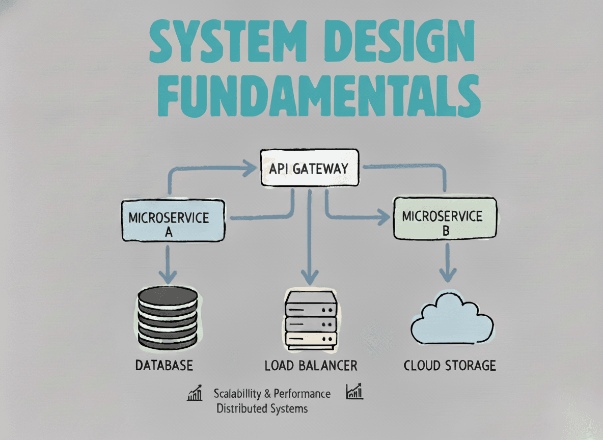
System Design Fundamentals
Build a strong foundation in designing scalable, reliable distributed systems.
View the courseSystem Design practice on Codemia
Work through 120+ system design problems with detailed solutions, from rate limiters to multi-region storage.